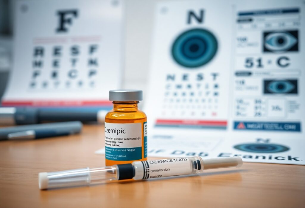
Vision can rarely be affected by Ozempic; you should monitor for sudden blurred vision or eye pain and seek care, while noting improved blood sugar and weight loss benefits. Understanding the potential Ozempic Side Effects is crucial for effective management.
Key Takeaways:
- Clinical trials (notably SUSTAIN-6) observed an increased rate of diabetic retinopathy complications with semaglutide, likely linked to rapid HbA1c lowering rather than direct eye toxicity.
- Patients with pre-existing diabetic retinopathy, long-standing or poorly controlled diabetes, or high baseline HbA1c carry higher risk of vision worsening after rapid glucose improvement.
- Reported ocular effects include blurred vision and other visual disturbances; true vision loss is uncommon but has been documented.
- Guidelines advise an eye exam before initiating semaglutide and closer ophthalmic monitoring for patients with active retinopathy; prompt reporting of new visual symptoms is recommended.
- Risk reduction strategies include aiming for gradual glycemic control, coordinating care with the diabetes team and an ophthalmologist, and individualizing treatment for those with severe retinopathy.
Understanding Ozempic and the GLP-1 Receptor Agonist Class
The Mechanism of Action: How Semaglutide Regulates Blood Sugar and Weight
Semaglutide activates GLP-1 receptors to boost insulin release and suppress glucagon, while slowing gastric emptying so you eat less; this lowers blood sugar and promotes weight loss, often producing noticeable appetite reduction and improved glycemic control.
The FDA Approval History and Primary Clinical Indications for Use
FDA approved semaglutide for type 2 diabetes in 2017 and a higher-dose formulation (Wegovy) for chronic weight management in 2021, so you can access it for those labeled indications under medical oversight and monitoring; official labels guide safe use.
Approval timelines reflect trial evidence showing consistent HbA1c reductions and meaningful weight loss, but you should follow prescribing guidance because dosing, contraindications, and monitoring differ between diabetes and weight-management indications, and off-label use can increase adverse-event risk.
Distinguishing Between Common Side Effects and Rare Adverse Events
Side effects like nausea, vomiting, or injection-site reactions are common and usually transient, while serious events such as pancreatitis or vision changes are rare; you should report new or worsening symptoms promptly.
Clinically, you should expect early GI symptoms that often improve, but uncommon severe events require urgent attention; if you have preexisting diabetic retinopathy you face a higher risk of vision worsening with rapid glycemic improvement, so baseline and follow-up eye exams are recommended and any visual changes must be assessed immediately.
The Emerging Connection Between Semaglutide and Ocular Health
Initial Case Reports and Early Clinical Observations of Vision Changes
Early case reports described patients who experienced sudden blurred vision after semaglutide, with a few instances suggesting a possible link to vision loss, prompting clinicians to alert you about monitoring visual changes.
Differentiating Between Transient Blurred Vision and Permanent Deficits
It is important to consider the Ozempic Side Effects when evaluating treatment options for diabetes and weight management.
Distinguishing transient blurriness from lasting damage helps you assess urgency: many episodes resolve with glucose or hydration shifts, while permanent deficits require immediate specialist care.
Assessment should include symptom timing, severity, pain, and your history of diabetic eye disease; bedside checks of visual acuity and pupillary responses can separate reversible metabolic or dry-eye causes from structural injury. If fundus exam or OCT demonstrates retinal or optic nerve pathology, you should pursue an urgent ophthalmic evaluation because permanent vision loss is the most dangerous outcome and demands rapid intervention.
The Role of Post-Marketing Surveillance in Identifying New Risks
Surveillance data and spontaneous reports have flagged ocular complaints after semaglutide, so you should report new visual symptoms and seek examination because post-marketing surveillance detects rare but serious events.
Systems such as national pharmacovigilance databases, EHR monitoring, and registries let you and clinicians submit adverse-event details while signal-detection tools seek patterns. When a safety signal emerges, regulators or manufacturers may fund studies or update labeling, meaning you gain earlier warnings and clearer guidance to reduce the chance of unrecognized new risks to your eyesight.
Non-Arteritic Anterior Ischemic Optic Neuropathy (NAION) Explained
Pathophysiology: What Happens to the Optic Nerve During an Ischemic Event
Ischemia deprives the optic nerve head of blood, causing axonal swelling and rapid loss of function. You may suffer sudden, often painless visual field defects as the swollen nerve compresses vulnerable fibers. Reduced perfusion and small optic discs are key risk factors that raise the chance of permanent damage.
Clinical Presentation: Identifying the Symptoms and Progression of NAION
You typically experience abrupt vision loss in one eye upon waking, commonly described as a shadow or blind spot; pain is usually absent. Symptoms often stabilize within days, but permanent central or peripheral deficits can remain, especially without prompt evaluation.
Visual field testing often shows an altitudinal defect and fundus exam reveals a swollen, pale optic disc in the acute phase; you may notice sudden worsening followed by relative stability. Fellow-eye involvement occurs in a minority of cases, so urgent vascular assessment and risk-factor control can reduce the chance of bilateral permanent loss.
Why NAION is Frequently Referred to as an “Eye Stroke” in Medical Literature
Clinicians describe NAION as an “eye stroke” because it stems from acute interruption of optic nerve blood flow, producing abrupt ischemic visual loss. You should note that mechanisms mirror cerebral ischemia even though the injury is localized to the optic nerve; rapid recognition matters because delay can mean permanent vision loss.
Pathology emphasizes small-vessel hypoperfusion rather than large-vessel embolism or inflammatory arteritis; you will not confuse NAION with arteritic AION without testing. Assessment focuses on visual fields, disc appearance, and systemic risk factors because early identification and management can limit bilateral involvement and long-term impairment.
Analyzing the Landmark Research: The Harvard/Mass Eye and Ear Study
Harvard and Mass Eye and Ear investigators analyzed large clinical datasets to flag signals linking Ozempic with eye problems; you should weigh the reported increased vision-loss risk against the study’s observational design and known limitations.
Study Methodology: Evaluating Observational Data and Patient Demographics
Researchers analyzed retrospective cohorts from electronic health records, matching by age, diabetes status and BMI; you should note the reliance on coded diagnoses, observational data, and remaining confounding risks.
Key Findings: Quantifying the Relative Risk Increase in Diabetic and Obese Populations
Results indicated a modest relative increase in vision-loss events among diabetic and obese GLP-1 users, while the study reported that absolute event rates remained low, which you must factor into clinical decisions.
Analysis of subgroup effects showed stronger relative estimates in people with long-standing diabetes and higher BMI, but the study lacked randomized allocation and had limited follow-up for chronic retinal outcomes. You should interpret elevated relative risks alongside the low absolute incidence, and consider that increased ophthalmic surveillance in treated patients could inflate observed cases; the authors’ sensitivity checks reduce but do not eliminate the possibility of residual confounding.
Critical Limitations: Understanding the Difference Between Association and Causality
Limitations include nonrandomized exposure, potential detection bias, and incomplete control for disease severity, so you must understand the findings show association not causation and cannot prove Ozempic directly causes vision loss.
Confounding arises when prescribing patterns differ by baseline risk and when vision outcomes are identified through billing codes rather than standardized ophthalmic adjudication. You should also account for surveillance bias, since treated patients may receive more eye exams, increasing case detection. Clinical trials with predefined, adjudicated retinal endpoints remain the only path to establish causality, so prioritize discussion with your clinician about risks and monitoring.
Biological Hypotheses: Why Ozempic Might Impact the Optic Nerve
Rapid Glycemic Control and Its Immediate Effect on Microvascular Stability
Rapid reductions in blood glucose from Ozempic can destabilize small retinal vessels, and you may notice transient worsening of diabetic retinopathy; watch for sudden vision changes and contact your clinician promptly if they occur.
The Presence of GLP-1 Receptors in Ocular Tissues and Potential Interactions
GLP-1 receptors are present in retinal and optic nerve cells, so Ozempic could interact directly with ocular tissue; you should be aware of a theoretical neurovascular modulation that might alter optic nerve function.
Evidence from animal and human tissue studies shows GLP-1 receptors in retinal ganglion cells and glia, and you should know receptor activation can modify intracellular signaling, axonal transport, and blood-retina barrier permeability; preclinical data suggest both protective and injurious effects depending on dose, timing, and metabolic context.
Systemic Vascular Changes: Blood Pressure Fluctuations and Ocular Perfusion Pressure
Fluctuations in systemic blood pressure after weight loss or medication adjustments can lower ocular perfusion pressure, and you might experience transient ischemic stress to the optic nerve; monitor blood pressure and report sustained drops or vision blurring.
You should understand that ocular perfusion equals arterial pressure minus intraocular pressure, so rapid systemic blood pressure falls can overwhelm autoregulation and cause optic nerve hypoperfusion; patients with glaucoma or advanced diabetic eye disease carry a higher risk of ischemic optic neuropathy, so coordinate changes with your eye care provider.
Risk Stratification: Identifying the Most Vulnerable Patient Profiles
Patients with long-standing diabetes, advanced age, or prior retinal disease carry the greatest concern; you should be prioritized for baseline retinal exams and closer follow-up, with pre-existing diabetic retinopathy marking the highest vulnerability.
The Impact of Pre-existing Ocular Conditions on Treatment Safety
If you have prior eye disease-glaucoma, macular edema, or proliferative retinopathy-your risk of visual deterioration while on semaglutide is higher, so schedule ophthalmic assessment before treatment and during early dose changes.
Dosage Correlation: Analyzing Risk Levels Across Different Strengths of Semaglutide
Lower semaglutide doses appear associated with fewer reported ocular events, but you still need monitoring; do not assume zero risk even at maintenance doses.
Higher doses and rapid upward titration have been linked in some reports to increased retinal complications; you should discuss dose selection with your clinician, monitor for new floaters, flashes, or blurred vision, and stop treatment pending evaluation if you notice any visual symptoms. Clinical data suggest a possible dose-dependent association, especially when combined with rapid weight loss or unstable glucose control.
The Intersection of Comorbidities: Sleep Apnea, Hypertension, and Vision Loss
Sleep apnea and uncontrolled hypertension amplify microvascular stress in the eye; you should keep blood pressure controlled and screen for sleep disorders to reduce the chance of vision loss while using Ozempic.
Combined obstructive sleep apnea, fluctuating hypertension, and chronic hyperglycemia create synergistic damage to retinal vessels; you should aim for steady glycemic and blood pressure targets, pursue CPAP for apnea when indicated, coordinate care between your prescriber and ophthalmologist, and seek urgent evaluation for any new visual changes to prevent permanent harm.
The Paradox of Diabetic Retinopathy and Rapid Glucose Lowering
Understanding the Temporary Worsening of Retinopathy During Intensive Therapy
Studies show that when you lower glucose rapidly, pre-existing diabetic retinopathy can worsen transiently, with the greatest risk in the first months, so you need prompt ophthalmic assessment and symptom vigilance.
Long-term Ocular Benefits of Glycemic Stability vs. Short-term Ischemic Risks
Evidence indicates that sustained glycemic control offers long-term protection against vision loss, while abrupt HbA1c drops can provoke short-term ischemic stress in vulnerable retinas.
Over months to years, you receive clear reduction in microvascular progression from steady glucose control, lowering lifetime blindness risk; Rapid reductions can induce retinal ischemia and edema, so you should balance pace of HbA1c decline with close retinal monitoring and treat active disease before intensifying therapy.
- Maintain steady targets when possible to maximize long-term benefit.
- Evaluate baseline retinopathy to assess short-term risk.
- Modulate rate of HbA1c reduction if advanced disease is present.
Benefits vs Risks
| Long-term Benefit | Short-term Risk |
|---|---|
| Reduced microvascular progression and lower lifetime blindness risk | Transient worsening, ischemia, macular edema |
| Improved overall ocular health with sustained control | Higher risk if proliferative disease is untreated |
Clinical Guidance for Managing Eye Health in Patients with Advanced Diabetes
If you have advanced or proliferative retinopathy, ensure a baseline dilated eye exam before rapid glucose lowering and arrange expedited ophthalmology follow-up.
Coordinate care with your diabetes team and an ophthalmologist so that active proliferative disease is treated (anti-VEGF or PRP) prior to aggressive HbA1c reduction; You should plan slower targets and scheduled ocular reassessments to reduce the chance of acute vision loss.
- Obtain baseline dilated exam before intensifying therapy.
- Refer to ophthalmology for severe or proliferative retinopathy.
- Schedule follow-up at 1-3 months after rapid glucose changes.
- Report visual changes immediately for urgent evaluation.
Clinical Actions
| Action | Rationale |
|---|---|
| Baseline dilated exam | Identifies risk and guides pace of glucose lowering |
| Treat active PDR | Reduces risk of hemorrhage and acute vision loss during therapy |
| Frequent post-change follow-up | Detects transient worsening early for timely intervention |
Warning Signs and Red Flags for Patients and Caregivers
Recognizing Sudden, Painless Vision Loss in One or Both Eyes
Watch for sudden, painless vision loss in one or both eyes; if you notice a curtain, sudden dimming, or a large blind spot without pain, contact emergency services or an ophthalmologist immediately.
The Importance of Monitoring Peripheral Vision and Visual Field Changes
Monitor peripheral vision by checking for missing side vision, difficulty spotting objects while driving, or narrowing fields; report any new blind spots to your eye doctor without delay.
Compare daily checks using an Amsler grid or simple spotting tasks and log any waviness, missing areas, or changes in size; bring those notes to appointments so your clinician can assess visual field deterioration promptly.
Immediate Action Steps: When to Contact an Ophthalmologist or Emergency Services
Contact emergency services for sudden vision loss or a curtain-like shadow and call your ophthalmologist the same day for new floaters, flashes, or progressive field loss; rapid evaluation can protect sight.
Prepare to report the exact time of onset, your Ozempic dose, and a current medication list when you call; avoid driving, arrange immediate transport, and follow triage instructions so clinicians can prioritize urgent care.
Medical Guidelines and Updated Prescribing Precautions
Guidelines now expect you to obtain a baseline ophthalmic assessment and to counsel patients about rare but serious vision loss risks when prescribing Ozempic, emphasizing early monitoring and prompt referral for any rapid visual changes.
New Recommendations for Baseline Eye Exams Before Starting Ozempic
Before you start Ozempic, you should order a baseline dilated eye exam, document retinopathy status, and schedule closer follow-up for those with preexisting diabetic retinopathy to reduce the chance of worsening retinopathy.
Informed Consent: How Physicians Should Discuss Rare Ocular Risks with Patients
You must include a clear discussion of rare ocular risks, symptom red flags, and instructions to seek urgent care, and document that conversation in the medical record.
When you obtain consent, describe specific symptoms like sudden vision loss, floaters, or flashes, explain that events are uncommon but potentially severe, record the discussion, and offer alternative treatments while stressing immediate ophthalmic evaluation for any change.
Interdisciplinary Communication Between Endocrinologists and Eye Specialists
Coordination between you and eye specialists should define expedited referral pathways, shared documentation of retinopathy staging, and agreed follow-up intervals to protect patient sight.
Regular communication via shared electronic records, direct urgent lines, and joint care plans lets you align on dose adjustments, monitor progression, and ensure rapid intervention when sight-threatening changes are suspected.
Regulatory Oversight and Potential Labeling Changes
The FDA’s Current Stance on Semaglutide and Optic Nerve Safety
FDA is monitoring reports of vision loss and optic nerve events with semaglutide without confirming causation; you should watch for visual symptoms and report them. Label updates remain possible if post‑market data show a clear risk.
European Medicines Agency (EMA) Investigations and Global Safety Updates
EMA opened investigations into reported optic neuropathy cases; you should expect regional safety alerts and coordinated data reviews. Regulators may request additional studies or temporary measures while assessments proceed.
European regulators have launched formal assessments after case reports suggested potential optic nerve injury; you will see periodic public updates and safety communications. The EMA can require targeted post‑authorization studies, compel labeling that includes explicit warnings about vision risks, and enforce risk‑minimization plans if evidence strengthens, with international agencies sharing data to speed conclusions.
The Impact of Clinical Findings on Future Product Liability and Safety Warnings
Legal consequences could follow if trials confirm a semaglutide-vision link; you might face increased lawsuits and heightened product liability claims. Manufacturers may add stricter warnings affecting prescribing and consent.
Manufacturers facing convincing clinical data are likely to revise labeling to include explicit optic nerve warnings, update patient leaflets, and mandate closer monitoring; you may be asked to acknowledge new risks. Courts often view stronger labels as evidence, which can raise settlement pressure and lead to higher out‑of‑pocket costs or insurer scrutiny for affected treatments.
Ozempic Side Effects – The Truth About Vision Loss Risk
The Role of Gradual Dose Titration in Minimizing Vascular Shock
Titration of Ozempic should proceed slowly so you and your clinician reduce the chance of abrupt hemodynamic shifts; this approach lowers the risk of vascular shock and sudden retinal perfusion decreases that can threaten vision.
Routine Monitoring Protocols for High-Risk Patients on Long-term Therapy
Schedule baseline and periodic ophthalmic exams if you have diabetes, hypertension, or prior retinopathy, and track visual acuity and new symptoms to catch early vision loss.
Monitor your status as a high-risk patient with a coordinated plan: arrange regular ophthalmology visits, report visual changes immediately, and aim for tighter glycemic and blood pressure targets; consider exams every 3-6 months while on therapy to detect progression of retinopathy or early signs of vision loss.
Managing Modifiable Risk Factors: Blood Pressure and Lipid Optimization
Optimize blood pressure and lipid therapy while you use Ozempic to reduce vascular strain and protect retinal perfusion; follow medication plans and lifestyle changes. Perceiving worsening blood pressure or cholesterol control should prompt you to intensify treatment and contact your clinician.
- Blood pressure
- Lipids
- Smoking cessation
Coordinate care to adjust antihypertensives and statins, monitor targets, and track your glycemic control to minimize vascular insults to the retina. Perceiving new visual symptoms or increasing variability in your blood pressure must trigger immediate reassessment and possible therapy changes.
- Antihypertensives
- Statins
- Glycemic control

Future Research Directions and Unanswered Questions
The Need for Prospective Clinical Trials to Confirm Causality
Prospective randomized trials are needed to determine whether GLP-1 agonists directly cause NAION; you should demand studies that measure incidence, timing, and dose-response, and include predefined ophthalmic endpoints to confirm causality.
Investigating Recovery Rates and Long-term Outcomes for NAION Patients
Recovery patterns after NAION on GLP-1 therapy remain unclear, so you must support longitudinal registries reporting visual acuity, field changes, and rates of permanent vision loss.
Longer-term follow-up should track visual acuity, automated perimetry, optical coherence tomography, vascular imaging, and systemic factors so you can identify temporal patterns, treatment effects, and predictors of recovery; emphasis on rates of permanent vision loss will clarify prognosis.
Exploring the Genetic Predispositions to GLP-1 Induced Ocular Changes
Genetic studies can reveal variants that increase your susceptibility to GLP-1-related ocular harm, enabling risk stratification and targeted monitoring for those with high genetic susceptibility.
Targeted genome-wide association studies and pharmacogenomic analyses should enroll diverse populations so you can detect common and rare variants linked to optic nerve ischemia; combining genetic risk scores with clinical factors could allow personalized risk stratification, inform decisions to avoid GLP-1 therapy in high-risk patients, and prompt early ophthalmic monitoring.
Conclusion
Hence you must discuss vision risk with your clinician; evidence links GLP-1 agonists like semaglutide to rare reports of vision changes. You should monitor for blurred vision or visual field loss and seek prompt ophthalmic evaluation if symptoms appear to balance treatment benefits against potential ocular harm.
FAQ
Q: Can Ozempic cause vision loss?
A: Clinical trials and post‑marketing reports do not show a direct toxic effect of semaglutide on the eye, but increased diabetic retinopathy complications were observed in the SUSTAIN‑6 trial (hazard ratio ~1.76) in patients with pre‑existing retinopathy and large, rapid falls in HbA1c. The product label includes a warning about diabetic retinopathy complications. Severe, permanent vision loss is uncommon, but cases of worsening retinopathy and vision changes have been reported. Discuss baseline ophthalmic assessment and monitoring with your prescriber if you have diabetes-related eye disease.
Q: How could Ozempic lead to worsening vision?
A: Rapid improvement in blood glucose can temporarily worsen diabetic retinopathy through changes in retinal blood flow, osmotic shifts, and progression of macular edema or proliferative disease. Semaglutide itself is not proven to damage retinal tissue directly; risk appears linked to the speed and magnitude of glucose lowering in patients with existing retinal disease. Treating active retinopathy before large glucose reductions can reduce the chance of vision complications.
Q: Who is at highest risk of vision problems while taking Ozempic?
A: Patients with pre‑existing diabetic retinopathy, long duration of diabetes, very high baseline HbA1c, or recent unstable glycemic control face the greatest risk. Prior history of proliferative retinopathy, macular edema, or recent retinal treatments further increases risk. Younger patients without retinopathy and those with well‑controlled diabetes have much lower risk.
Q: What vision symptoms should prompt immediate medical attention?
A: Seek urgent ophthalmic care for sudden loss of vision, new or rapidly increasing floaters, flashes of light, or a sudden increase in blurriness or distortion. Contact your diabetes clinician promptly if these symptoms occur so they can coordinate care with an eye specialist and consider temporarily adjusting therapy. Early evaluation and treatment of retinal bleeding or macular edema improves outcomes.
Q: How can patients and clinicians reduce the risk of retinopathy complications when using Ozempic?
A: Obtain a baseline dilated eye exam before starting semaglutide if you have long‑standing diabetes or known retinopathy and schedule follow‑up exams as recommended by your ophthalmologist. Aim for controlled, paced HbA1c reduction rather than very rapid large drops when retinal disease is present, and coordinate systemic glucose management with retinal treatment (laser or intravitreal therapy) when needed. Report any visual changes immediately; your clinician may modify therapy or refer for urgent retinal care.